U-arm (graphy, scopy)
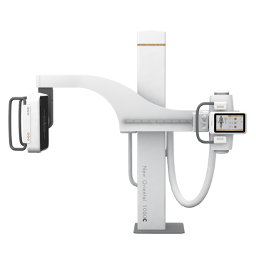
U-arm with a flat panel detector is a universal digital system for clinics and hospitals, private and public, with a high flow of patients. The system can meet the needs of most common examinations, both vertical and horizontal, including breast examinations.
Faster, Lighter, Safer - the filmless process will be more convenient and faster than the conventional radiographic process. Digital radiology significantly reduces diagnostic time, simplifies the examination process and facilitates relationships between patients and radiologists. U-arm with flat panel detector DICOM compatible.
Compact design. One of the unique features of the FPD DR U-handle system is its compact mechanical design. Its installation site requirements are the least demanding among various types of digital radiography systems, making it an ideal choice for small installation sites. Typically, a typical 15 m2 room will adequately accommodate the entire system, including the isolated control panel.
Peculiarities:
Fully digital system
Dynamic Flat Panel Detector and Tube from Siemens, Provides Amazing Images
Touch control panel for tube and detector
Unique design
|
Name |
Specifications |
| High frequency generator | Catalog number: GFS501-1 Power: 65kW Radiography kV: 40~150Kv; mA Range: 10~800mA Fluoroscopy kV: 40~125kV, Fluoro mA: 10~40mA Exposure time: 1ms~ 10s, mAs: 0.1mAs~ 1000mAs Preset programs: 600 positions, Automatic exposure control Power: 380VAC, three-phase Network integration: generator parameter control and image acquisition are integrated on a single platform. Detection and diagnosis of faults. |
| Dynamic Flat Panel Detector |
|
| X-ray tube assembly SIEMENS |
|
| U-shaped structure | Catalog number: SZ-9 Vertical movement range (U-arm in horizontal position): 1300 mm Minimum distance between focus and ground (U-arm in horizontal position): ≤450mm Motorized Rotation Range (U-arm): (-30°~ 0°~ +90°~ +120°) Detector rotation range (motorized): -45°~ +45° SID: 1000mm~1800mm, motorized |
| Aluminum Interdimensional Lattice | Mesh ratio: ≥8:1 Reticle focus: ≥130cm |
|
Collimator |
Catalog number: XS2-7 Radiation field (SID = 1000 mm): max.: 430 mm × 430 mm; Min:0mm×0mm Operating mode: manual |
| Radiographic table | Catalog number: SC1-2 Length: 1950 mm; Width: 680 mm; Height: 680 mm Attenuation equivalent <1.2 mm Al Foot wheel brake: >100N Load capacity: 200 kg |
| Remote control | Remote control of mechanical system movement |
| Imaging workstation | Hardware Configurations: Processor: Intel Core 2 Duo (Core 2 Duo) ≥3.7 GHz Memory: 4 GB; Hard Drive: ≥1TB DVD R/W System interface: USB, 1000 MB network interface, DVI/HDMI monitor 24-inch LCD monitor (color); Resolution: 1920×1080 OS: Windows 7 or later Software Configuration: WD-ACQUIRE Image acquisition: setting acquisition conditions, setting mechanical position, APR Fluoro data acquisition: 25 fps, 15 fps, 4 fps, 2 fps, 1 fps Data management: patient registration, patient information and image management, report management Recording and displaying exposure data DICOM Service: Send, Receive, Store, Query, WORKLIST, MPPS, Validate, etc. DICOM SR: reporting, storage, printing, sending Data statistics: statistics of counting exposure, body part, number of patients, etc. for a specific date or period. RIS support. Image processing: window width/level, automatic window width/level adjustment, preview, preset window width/level, inverting positive and negative images; The image is flipped, rotated, scaled, moved; Image interpolation edge enhancement, original image annotation restoration, character/number annotation, image annotation, tape measurement, area measurement, auto-tuning. Image printing: DICOM printing, paper printing, manual selection of images displayed for printing, one-key annotated image printing, compatible with various printing equipment, film format printing, number of prints, print queue management, stop/start preset. Image enhancement filter: According to the different physiological structure of body parts and different diagnostic requests of doctors, and different clinical requirements, algorithms are optimized for different parts of the body. Personal settings: format and layout, default settings, toolbar settings. Other Features: Users can define the display format or layout; You can use different methods to view patient images and determine how to find information. Redundancy function, pixel optimization, diagnostic workstation image processing software interface in English, support high-speed lossless compression transmission, support online decompression. The operating interface is integrated with generator control and mechanical system status display, allowing all generator parameters such as kV exposure, mA, mAs, exposure time, ionization chamber activated field, APR, etc. to be configured on the operating interface. Exposure status and error code. |
 Catalog number: WDF 4343R
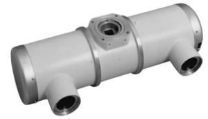 Catalog number: RAY-14_1