Digital Ultrasound Diagnostic System Acclarix LX8
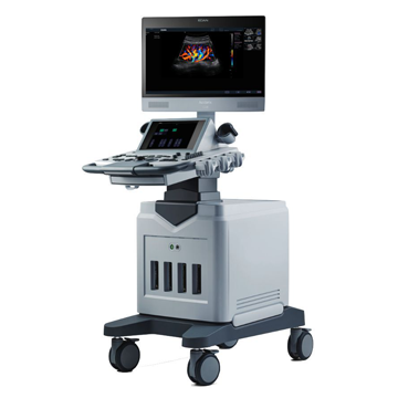
The new device from EDAN belongs to a new generation of ultrasound scanners. The Acclarix LX8 is designed from scratch with a focus on delivering cutting-edge levels of innovation and performance at a price that surprises.
System Features:
• 21" high-definition monitor with tempered glass coating
• 10" touch screen with customizable user interface
• Motorized height adjustment
• 4 active ports
The EDAN Acclarix LX8 ultrasound scanner provides excellent image quality for ultrasound diagnostics in various areas:
• Abdominal examinations of adult and pediatric patients
• Obstetrics and gynecology, incl. 3D/4D
• Cardiology
• Vascular examination
• Examination of superficial organs
• Research of the musculoskeletal system
• Neonatology
• Urology
Specifications:
• Imaging modes: B, B/B, 4B, B/M, M
• CW - Continuous Wave Doppler
• PW (Pulse Wave Doppler) - Pulse Wave Doppler
• CDFI (Color Doppler Flow Imaging)
• PDI (Power Doppler Imaging) - Power Doppler
• Directional PDI (DPDI) – Directional Power Doppler
• Pulse Inversion Harmonic - tissue inversion harmonic
• 4D volumetric visualization mode in real time
• 3D volumetric reconstruction mode
• Panoramic scanning mode
• Trapezoidal scanning
• 21-inch LCD monitor with high resolution
• 10 inch touch control interface
• Electric console height adjustment
• Connector for connecting 4 sensors
• THI - Tissue Harmonica
• TSI - Technology for recognizing the specificity of the tissues being studied
• Scanning depth (mm): up to 300
• Cine loop up to 12287 frames
• Internal archive for storing images (archiving images and cine loops to the built-in 1 TB hard drive or USB)
Scanning and image processing technologies:
- Multi-beam imaging (mBeam)
- Image Formation Optimization (BFO)
- SpeckleResistance (eClear) (image grain suppression)
- Phased Inversion Harmonic Compound Imaging (ultrasound beam boosting mode (eHCI)
• Multilingual interface (Russian, English, German, French, Chinese)
• Software for research: abdominal, obstetric, gynecological, small organs, pediatrics, cardiology, urology, vascular, thyroid and mammary glands
• Auto Doppler spectrum tracing with auto measurements
• Built-in ports: VGA -1 pc., Video -1 pc., USB -6 pcs., DICOM 3.0 -1 pc., (optional), Ethernet port, printer control port, pedal connection port
• Power supply: 220 V mains operation
• Dimensions, cm: 100 X 70 X 140-180 (L x W x H)
• Weight: 90 kg
Sensors:
• Convex sensor C5-2XD (2-5 MHz / H2-5 MHz)
• Linear sensor L10-4D (4-9 MHz / H5-10 MHz)
• Endocavital sensor E8-4D (4-8 MHz / H5-8 MHz)
• Sector phased sensor P5-1XD (1-5 MHz / H2-5 MHz)
• Volume convex sensor C5-2MD (2-5 MHz / H3-5 MHz)
• Linear sensor L12-5D (5-11 MHz / H6-12 MHz)
• Linear sensor L17-7HD (7-15 MHz / H9-17 MHz)
• Linear sensor L17-7SD (7-15 MHz / H9-17 MHz)
• Microconvex sensor MC8‐4D (4-8 MHz / H4-8 MHz)