Portable Ultrasound Diagnostic System U50
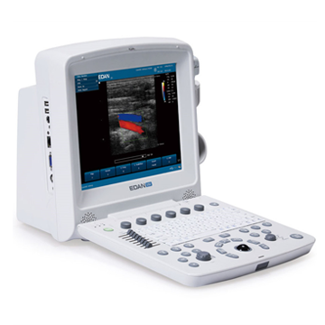
Currently, the Edan U 50 ultrasound scanner is one of the most popular models on the global ultrasound scanner market. The manufacturing company doubled the number of transmitting channels, which led to a significant increase in the quality of the resulting image, graininess and noise decreased. EDAN U50 is the optimal ultrasound system for a newly opened medical center, for a small ultrasound room, or for those who want to move from screening monochrome ultrasound scanners to a new level of diagnostics.
• Ultrasound scanner EDAN U 50 can also be used in veterinary medicine. It has special veterinary programs.
Basic information about the EDAN U50 ultrasound scanner
• CW Doppler
• Portable device with color Doppler.
• The most affordable price in this class of ultrasound scanners.
• Modes: B, B/B, 4B, B/M, M, PDI, PWD, CDFI,
• Tissue inverse harmonic,
• Tissue harmonics,
• Scanning depth up to 320mm
• LED monitor 12.1 inches,
• Connection of 2 sensors,
• Possibility of battery operation,
• Weight 7.1kg.
Main functions of EDAN U50
• Imaging modes: B, B/B, 4B, B/M, M
• CWD - Continuous Wave Doppler
• PDI (Power Doppler Imaging) - Power Doppler
• PWD (Pulse Wave Doppler) - Pulse Wave Doppler
• CDFI (Color Doppler Flow Imaging-) Color Doppler
• Pulse Inversion Harmonic - tissue inverse harmonic
• 12.1-inch high resolution LED monitor
• Simultaneous connection of 2 sensors
• THI - Tissue Harmonic
• TSI - Technology for recognizing the specificity of the tissues being studied
• 8-segment TGC correction
• 16-segment acoustic output power control
• Scanning angle: from 30 to 155 degrees
• Scanning depth (mm): from 20 to 300
• Real-time zoom: x1.0 x1.2 x1.4 x1.6 x2.0 x2.4 x3.0 x4.0
• Cine loop for 256 frames
• Built-in memory for storing images
• Built-in battery (optional)
Technologies used in scanning and image processing:
-Multi-beam imaging (mBeam)
- Image Formation Optimization (BFO)
-Speckle Resistance (eClear)
-Phased Inversion Harmonic Compound Imaging (ultrasonic beam swing mode) (eHCI)
• Russian-language menu
• Russified software for research: abdominal, obstetric, gynecological, small organs, pediatrics, cardiology, urology, blood vessels, thyroid and mammary glands
• Built-in ports: VGA -1 pc., Video -1 pc., USB -2 pcs., DICOM 3.0 -1 pc., (optional), Ethernet port, printer control port, pedal connection port
• Power supply: Operation from 220 V mains and from battery.
• Dimensions (width x height x length): 320mm X 330mm X 220mm
• Weight 7.1kg
Modes of the ultrasonic scanner EDAN U 50
• Pulse Inversion Harmonic (tissue inversion harmonic) - technology for isolating the harmonic component of vibrations of internal organs caused by the passage of basic and inverse ultrasonic pulses through the body. The signal obtained by adding the base and inverse components of the reflected signal is considered useful. Typically, an inverse harmonic (compared to a direct harmonic) provides better quality because both signals (base and inverse) pass through the body and noise is automatically filtered out when added. It is most appropriate to use inverse harmonic technology when studying moving tissues (vessels, heart) and difficult-to-visualize tissues (with similar acoustic density), such as tumors.
• Power doppler (power doppler) - qualitative assessment of low-speed blood flow, used in the study of a network of small vessels (thyroid gland, kidneys, ovary), veins (liver, testicles), etc. More sensitive to the presence of blood flow than color doppler. The echogram is usually displayed in an orange palette; brighter shades indicate a higher blood flow rate.
• Pulse Wave Doppler (pulse wave Doppler) - used to quantify blood flow in blood vessels. The vertical time base displays the flow velocity at the point under study. Flows that move towards the sensor are displayed above the baseline, reverse blood flow (from the sensor) is below. The maximum flow speed depends on the scanning depth, pulse frequency and has a limitation (about 2.5 m/s when diagnosing the heart). High-frequency pulsed Doppler (HFPW - high frequency pulsed wave) allows you to record higher flow velocities, but also has a limitation associated with distortion of the Doppler spectrum.
• Color Doppler Flow Imaging - highlighting the nature of blood flow in the area of interest on the echogram in color (color mapping). The blood flow to the sensor is usually mapped in red, and from the sensor - in blue. Turbulent blood flow is mapped in blue-green-yellow color. Color Doppler is used to study blood flow in vessels and in echocardiography. Other names for the technology are color Doppler mapping (CDC), color flow mapping (CFM) and color flow angiography (CFA).
• Continuous Wave Doppler or CW (Continuous Wave Doppler) is used to quantify blood flow in vessels with high-speed flows. The disadvantage of the method is that the flows are recorded integrally, i.e. throughout the entire scanning depth. In echocardiography, using continuous Doppler, it is possible to calculate the pressure in the cavities of the heart and great vessels in one or another phase of the cardiac cycle, calculate the degree of significance of stenosis, etc. The basic equation for CW is the Bernoulli equation, which allows the pressure difference to be calculated. Using this equation, you can measure the pressure difference between the chambers under normal conditions and in the presence of pathological high-velocity blood flow.
• Tissue Harmonic Imaging (THI, tissue or 2nd harmonic) - technology for isolating the harmonic component of vibrations of internal organs caused by the passage of a basic ultrasound pulse through the body. The useful signal is the one obtained by subtracting the base component from the reflected signal. The use of the 2nd harmonic is advisable when ultrasound scanning through tissues that intensively absorb the 1st (basic) harmonic. This technology involves the use of broadband sensors and a high-sensitivity receiving path. Improves image quality, linear and contrast resolution in overweight patients.