Optical coherence tomograph (anterior chamber) CASIA 2
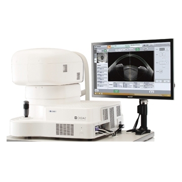
CASIA2 offers intuitive and largely automated control and incredible measurement speed.
CATARACT
An analysis of the anterior and posterior surfaces of the cornea will help determine the best refractive procedure for patients;
A unique measurement of all lens parameters allows you to accurately predict the outcome of the operation;
Postoperative screening clearly visualizes and documents the quality of the treatment outcome.
GLAUCOMA
Automatically detects the anterior camera angle at 360 degrees and displays the result in a detailed and three-dimensional map;
Various scanning methods allow you to visualize tiny areas of interest and provide amazing scanning accuracy.
CORNEA
All measurements of different areas of the cornea can be displayed on a clearly organized corneal analysis map;
Unique trend analysis displays chronological progress after treatment;
In addition to the Ectasia software, a new epithelial map is now available, so that all changes in the cornea will be immediately detected.
MEASURING UNIT
Resolution - axial depth - 10 µm or less (in tissue), transverse - 30 µm or less (in tissue)
Scanning speed - 50,000 A-scans/sec
Scanning range - 16x16x13 mm
Transverse raster - 12x12 mm
Measuring head travel - 12x12 mm, 88 mm (X-axis), 40 mm (Y-axis), 43 mm (Z-axis)
Chin rest adjustment - 70 mm
Touch screen - 20’’
Dimensions (d/w/h) - 530x560x455mm
Weight - 33 kg
Setting mode - manual, joystick, touch screen, auto-tuning, auto shots
Light source type - SWEPT source
Wavelength - 1,310 nm
Principle - Fourier Domain
Output power - Less than 6mV
POWER SUPPLY
Voltage - 100V-240V
Frequency - 50/60Hz
Power consumption - 170VA
OS - Windows 8.1 64 bit
Processor - Intel Core i7 or higher
Memory - 8 GB
SSD or HDD - SSD 128 GB and external 2-8 TB
Data export - Printer (LAN/USB)
Data import - LAN/USB
MEASUREMENT MODES
Scanning method - Radial/raster/2D H+V/2D single/Film H+V
Depth - 11 mm and 13 mm
B-scan width - 3 mm-16 mm
A/V scan - 400-2000 A-scans per line
V/C scan - 8/16/32/64/128/256
Fixation - point/accommodation/periphery
Setting - auto/manual/Z-off
Cornea map
Scan direction - radial scan - 16 images
Resolution - 800 A-scans per line
Scanning speed - 0.3 sec
Scanning range - Ø 16 mm
Depth - 11 mm
Filtration pad
Scanning direction - raster scanning - horizontal, vertical, 256 images
Resolution - 400 A-scans per line
Scanning speed - 2.4 sec
Scanning range - B12xC12 mm
Depth - 11 mm
Global AS Scan
Scanning direction - radial scanning - 128 images
Resolution - 800 A-scans per line
Scanning speed - 2.4 sec
Scanning range - Ø 16 mm
Depth - 11 mm
Lens biometrics
Scan direction - radial scan - 16 images
Resolution - 800 A-scans per line
Scanning speed - 0.3 sec.
Scanning range - Ø 16 mm
Depth - 13 mm
Vitreous body raster
Scanning direction - raster-256 images
Resolution - 400 A-scans per line
Scanning speed - 2.4 sec.
Scanning range - B12xC12 mm
Depth - 13 mm
Global lens scan
Scanning direction - radial scanning - 128 images
Resolution - 800 A-scans per line
Scanning speed - 2.4 sec.
Scanning range - Ø 16 mm
Depth - 13 mm
Angle analysis
Scanning direction - radial scanning -16 images
Resolution - 800 A-scans per line
Scanning speed - 0.3 sec
Scanning range - Ø 16 mm
Depth - 11 mm
Angle in HD image
Scanning direction - radial scanning - 64 images
Resolution - 800 A-scans per line
Scanning speed - 1.2 sec
Scanning range - B8xC4
3D/2D analysis
3D overview - gonioscopy, slices, rotations, ITC
Maps - Axial force (anterior, posterior, real), Refractive power (keratometric, anterior), Instantaneous force (keratometric, anterior, posterior, real), Elevation (posterior, anterior), pachymetry (map, sectors), Epithelium, Anterior depth cameras, OCT (horizontal, vertical)
Analysis function - Axial force (anterior, posterior, real), Refractive power (keratometric, anterior), Instantaneous force (keratometric, anterior, posterior, real), Elevation (posterior, anterior), pachymetry (map, sectors), Epithelium, Depth anterior chamber, OCT (horizontal, vertical)
Export video - View in 2D rotation mode/C-scan -3D video