Floor (graphy)
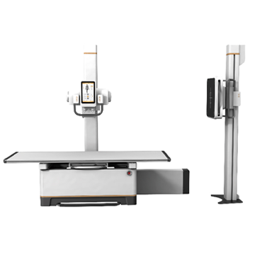
A universal two-person digital system for clinics and hospitals, private and public, with a high flow of patients. The system can meet the needs of most common examinations, both vertical and horizontal, including breast examinations. Actual contents and features vary depending on the configuration selected.
It is a combination of digital flat panel imaging and classical radiographic mechanical design. The system is suitable for most common clinical radiography applications in both vertical and horizontal projections, as well as chest examinations, significantly increasing patient throughput.
Of course, your film-free workflow will be more convenient and faster than a conventional radiographic process. Patients also benefit from low radiation levels and smooth procedures.
Complies with DICOM 3.0 standard, which means you can benefit from all relevant DICOM services, including DICOM worklist, image storage, printing and other features that will greatly improve your workflow.
The high resolution a-Si flat panel detector provides high image quality, the a-Si FPD detector technology marks the highest level of X-ray imaging technology.
The detector's superior detection quantum efficiency (DQE) allows for more image detail to be revealed with less radiation dose. The 9.2 megapixel resolution makes the image perfect.
The diagnostic procedure is greatly facilitated by the ideal size of the detector (17" x 17"), since any part of the body can be examined in any position, and thus inconvenient positioning work is significantly reduced.
Classic mechanical structure
The combination of a floating x-ray table, tube stand and vertical support post is the most popular and practical x-ray room configuration.
Advanced digital data acquisition and processing system InvaRay
Based on our InvaRay digital imaging platform, several new WDM patented technologies are integrated. In addition to further improving image quality, our customers are also offered a number of innovative clinical imaging features.
Multispectral processing method
The MCO function can divide image signals into multiple channels according to their distinctive features. The signals in each channel are then calibrated and adjusted according to their respective characteristics and the different focus of each image.
In this way, all the details of the image can be best displayed.
Automatic shutter
The afterimage of the collimator has been eliminated; This way, the visual impact of the image is optimized, ensuring that the ROI is most noticeable. Meanwhile, comprehensive post-processing tools provide radiologists with ample space for image calibration and processing.
Auto APR (Anatomical Program)
The modern Auto APR function allows you to automatically set parameters for examining different parts of the body and different positions. It can also automatically select suitable post-processing methods to optimize and characterize image quality. All work can be completed by pressing the manual switch with optimal image output.
Digital correlation
A new digital correlation algorithm provides precise image stitching, allowing easy imaging of the entire spine and legs.
Convenient one-button data retrieval function
Based on the InvaRay software platform, compatible with the DICOM worklist. After receiving the necessary patient information from the HIS/RIS system before surgery, the system will automatically complete the parameter settings. At the same time, personalized recommendations for imaging parameters are provided that are better tailored to the viewing habits of individual surgeons, freeing them from complex surgical equipment settings and allowing them to focus on the operation.
Dedicated image acquisition protocol
The dedicated acquisition protocol incorporates filtering for specific body parts and gray balance processing into the automatic image optimization, display function, and image acquisition process. Based on years of image processing and clinical experience, WDM has built in hundreds of acquisition curves to clearly demonstrate the image of even a small focus.
Comprehensive service provides hospitals with a complete digital solution
WDM has been involved in the digital development of radiography for many years; thus representing a fully digital solution for hospitals rather than just digital equipment. With the convenience provided by WDM, you can create and expand your own RIS, PACS and HIS systems.
Peculiarities:
• Adjusting the height of the tube with the stand
• Canon X-ray tube (Japan)
• Automatic exposure control
• Radiation dose indicator (optional)
• Flat panel detector size 43x43 (wireless – option)
• Multispectral image processing
• WD Acquire branded workstation
• Panoramic X-ray function (optional)
|
Name |
Specifications |
| High frequency generator | Catalog number: GFS501-1 Power: 50kW Radiography kV: 40~150Kv; mA Range: 25~630mA Exposure time: 1ms~6.3s, mAs: 0.4mAs~630mAs Preset programs: 600 positions, Automatic exposure control Power: 380VAC, three-phase Detection and diagnosis of faults. Network integration: thanks to intelligent CAN-BUS communication system generator control is integrated with image acquisition on a common platform. The parameters and status of the generator can be displayed and monitored through the image acquisition system. |
| Flat Panel Detector | Catalog number: WDF 4343RP Detector type: a-Si Effective area: 430mm×430mm (17” ×17”) Weight: 5.5kg Matrix (pixel): 3028 ×3028, (9.4 megapixels), Pixel size: 142um A/D Converter: 16bit Spatial resolution: 3.6 lp/mm Image reconstruction time: <5s DQE: ≥70% MTF: ≥58% Ambient temperature: 10℃~40℃, Storage temperature: -15℃~55℃ Data transmission: Giga Ethernet Transmission of control commands: Giga Ethernet Calibration mode: offset calibration, gain calibration, error pixel calibration, line noise calibration Internal trigger control mode, applicable to upgrade the general system |
| X-ray tube assembly Toshiba | Catalog number: E7843X Focal spot: 0.6mm/1.2mm Power: 22/50kW Heat capacity of anode: 150kHU Target angle: 12° |
| Collimator | Catalog number: XS1-4 Radiation field (SID = 1000 mm): max.: 430 mm × 430 mm; Min:0mm×0mm Operating mode: Manual |
|
Table & Buka stand |
X-ray table Catalog number: SC4-5 Height from table top to floor: ≤ 670 mm Tabletop movement range (floating) longitudinal ≥ 905 mm, transverse ≥ 260 mm Longitudinal tube movement: ≥ 2400 mm X-ray tube vertical movement (tube focus on floor): 525mm~1760mm (Motorized) X-ray tube rotation along the horizontal arm: +120°~-120° Tube column rotation: 0°~±180° (fixed at ±90°, 0° and 180°) Longitudinal movement in relation to Beech post ≥ 540 mm Tabletop weakening equivalent <1.2 mm Al Load capacity: 250 kg LCD touch screen: 10.1 inches Beech Stance: Catalog number: LS-6 Detector vertical movement range (from center to floor): 370mm ~ 1800mm Automatic tracking of the X-ray tube to the detector level |
| Lattice | Mesh ratio: ≥8:1, f0: ≥100cm, aluminum based |
| Imaging workstation | Hardware Configurations: Processor: Intel Core 2 Duo (Core 2 Duo) ≥3.7 GHz Memory: 4 GB; Hard Drive: ≥1TB DVD R/W System interface: USB, network interface 1000 MB, DVI/HDMI monitor 24-inch LCD monitor (color or monochrome); Resolution: 1920×1080 OS: Windows 7 or later Software Configuration: WD-ACQUIRE Image acquisition: setting acquisition conditions, setting mechanical position, APR Data management: patient registration, patient information and image management, report management Recording and display of exposure data DICOM Service: Send, Receive, Store, Query, WORKLIST, MPPS, Validate, etc. DICOM SR: reporting, storage, printing, sending Data statistics: statistics of counting exposure, body part, number of patients, etc. for a specific date or period. RIS support. Image processing: window width/level, automatic window width/level adjustment, preview, preset window width/level, inverting positive and negative images; The image is flipped, rotated, scaled, moved; Image interpolation edge enhancement, original image annotation restoration, character/number annotation, image annotation, tape measurement, area measurement, automatic adjustment. Image printing: DICOM printing, paper printing, manual selection of images displayed for printing, one-key annotated image printing, compatible with various printing equipment, film format printing, number of prints, print queue management, stop/start preset. Image enhancement filter: According to the different physiological structure of body parts and different diagnostic requests of doctors, and different clinical requirements, algorithms are optimized for different parts of the body. Personal settings: format and layout, default settings, toolbar settings. Other Features: Users can define the display format or layout; You can use different methods to view patient images and determine how to find information. Redundancy function, pixel optimization, diagnostic workstation image processing software interface in English, support high-speed lossless compression transmission, support online decompression. The operating interface is integrated with generator control and mechanical system status display, allowing all generator parameters such as kV exposure, mA, mAs, exposure time, ionization chamber activated field, APR, etc. to be configured on the operating interface. Exposure status and error code. |