Multi-slice Computer Tomograph 32 slice
I. Configurations: 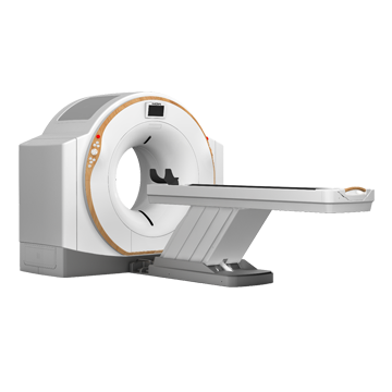
Hardware:
Name Model Quantity
X-ray system Generator KTHVG X4201 1
X-ray tube GS-30729 1
Collimator CTXS-2 1
Imaging System Imaging Software WD-CT Purchase 1
Detector WDDAS-1 1
Monitor 24" color LCD 1
Integrated Workstation Professional Workstation 1
Other equipment Portal SMZh-4 1
Patient table SMK-2A 1
KT console CTBOX-2 1
Power distribution PSB-10 1
II. Data sheet:
Detector Scintillation material GOS rare earth ceramic
Physical line number 24 rows
16mm Z-axis submillimeter coating
Number of detector blocks in a row 984
Minimum size of the detector unit along the Z axis 0.5 mm
Tube and generator Anode target surface Rhenium tungsten alloy
Thermal capacity of the tube is 3.5MHU
Tube cooling method Oil cooling and wind cooling
High voltage generator power 42kW
Optional tube voltage range 70kV, 80kV, 100kV, 120kV, 140kV
Maximum lamp output current 350 mA
Minimum tube output current 10 mA
Focus tube (small, large) 0.7 mm × 0.8 mm
1.2 mm × 1.4 mm
Gantry Type Slip Ring Low Voltage Slip Ring
Road Leather Drive Belt
Data transmission mode Radio frequency and optical fiber
Scan frame aperture 760 mm
Digital tilt Scan frame angle Digital ±30°
3D laser positioning system Yes
Portal cooling mode Wind cooling
Portal control panel Yes
Graphic breathing guidance Yes
Voice prompts for breathing control Yes
Table Maximum range of movement 2100 mm
Maximum scanning range in one spiral 1620 mm (optional 1750 mm)
Positioning image length 1620 mm (optional 1750 mm)
Check bed positioning accuracy ±0.25mm
Foot switch table control Yes
Maximum table load capacity 210 kg
Table movement control Manual and motorized
Scan parameters Scan layer/360° 32 image slices/360°
Fastest scanning time /360° 0.5 s/360°
Thin image scanning layer thickness 0.5mm
The thinnest image reconstruction layer is the thickest. 0.5 mm
Step range 0.15-2.0
Free choice of site Yes
Scan mode Scout image, axial, spiral
Maximum image acquisition matrix 1024×1024
Maximum image restoration matrix 1024×1024
Maximum image display matrix 1024×1024
Scanning time for one spiral 100 s
Spatial resolution 18LP/cm
Density resolution 2 mm at 0.3%
Integrated workstation
(Shooting + post-processing)
Display screen size 24 inches
Memory 32 g
Hard drive 2T
CPU
Processor 6 cores
Image format and transmission storage: DICOM 3.0 standard. Yes
Synchronous Parallel Image Processing Function Yes
Remote Control Service Diagnostic Interface Yes
Clinical Functional Software Low Dose Lung Scanning Technology Yes
Low dose pediatric scanning technology Yes
Intelligent tube current automatic control technology Yes
Iterative noise reduction method Yes
Beam Hardening Artifact Correction Software Yes
Posterior fossa image optimization Yes
Metal Artifact Removal Technique Yes
3D reconstruction module Yes
Maximum Projection Density MIP Yes
Minimum Density Projection MinIP Yes
Average Density Forecast AIP Yes
Intervertebral disc reconstruction module Yes
Report module Yes
Print module Yes
Automatic dialing and printing Yes