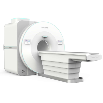
Magnetic Resonance Tomograph 1.5T Helium-free
There is no possibility of helium loss during quench
Automatic recovery and preservation of functionality
There is no need to refill helium every 3-5 years and during quench.
No more additional costs.
Magnet
1.5T MRI without liquid helium uses innovative ultra-low temperature conductive technology, integrates high-efficiency low-temperature environment, ultra-high vacuum, super insulation technology, aerospace grade carbon fiber suspension and other black technologies, truly realizes 100% magnetic resonance, and usheres in a new era of magnetic resonance without liquid helium.
Then there is no need to equip a superconducting MRI quenching tube, no need to supply liquid helium, and no need to be afraid of quenching. This method will stimulate future MRI examinations, significantly reduce costs, and benefit all of humanity.
To take into account magnetic field uniformity and patient comfort, the superconducting magnet has been professionally designed and calculated. While magnetic field uniformity has reached the best level in the industry, it also reduces patient claustrophobia to the greatest extent and provides a hardware platform for superior clinical images and advanced imaging functions.
Field strength: 1.5 T;
Aperture diameter: 60cm
Magnet length: <1600mm
Shielding method: active + passive
Magnetic field stability: ≤0.10 ppm/h
Uniformity: 40 cm DSV Vrms ≤ 0.2 ppm, 50 cm DSV Vrms ≤ 1.0 ppm
5G field: ≤ 4 m (axial), ≤ 2.5 m (radial)
Weight: about 4000 kg
Shimming: active + passive + dynamic
Patient table
A patient table is used to position the patient with associated RF receiving coils relative to the isocenter of the magnet for MRI scanning.
The patient table unit consists of two parts: the patient table and the positioning system.
The fully automatic patient table is easy to access and position.
The laser positioning system combined with the LCD display helps the doctor position the patient quickly and easily.
Max. patient weight: 250 kg
Tabletop longitudinal movement range: ≥ 2000 mm
Tabletop vertical movement range: ≥ 350 mm
Positioning accuracy: ±1mm
Spectrometer
The spectrometer is the most basic, high-tech component of an MRI system. Spectrometer R&D capabilities are the most important R&D capabilities of an MRI manufacturer. Typically only an in-house spectrometer is used, and WDM also has in-house spectrometers from 8 to 24 channels for superconducting MRI systems. Using the most advanced electronic technologies. It is equipped with all-digital RF transmission and reception technology; LVDS high-speed serial transmission and FMC high-speed data transmission technology; MRI-compatible chips with low power consumption and high SNR; High-precision sequence manager, proprietary homologous phase coherence technology, transmitting and receiving modules using the same local oscillator have good phase consistency and can remove frequency rewinding. Its advantages are stable performance, convenient use, easy operation, high speed and high image quality.
Receiving channel: 16-32 channels, and FMC technology allows you to expand the channels freely;
Modulation method: frequency, phase, amplitude
RF Frequency and Phase Accuracy: 32bit and 16bit;
RF and Gradient Wave Update Rate: 1US;
Gradient output channel: x, y, z and B0;
Gradient pre-extraction: 12 groups of straight and cross elements for eddy current compensation;
Homologous phase coherence technology: phase fluctuation ≤ 0.01°;
Digital dynamic range enhancement technology: effective bit 32 bits;
Fiber optic technology: fiber optic imaging, fiber optic gradient, no interference, low attenuation, easy connection and stable operation.
Gradient system
The gradient system is a modern system with active shielding and water cooling.
Independent water-cooled primary and secondary coils ensure high performance and low power consumption.
Gradient strength: 35 mT/m (single-axis)
Slew rate: 175 mT/m/ms
Minimum climb time: ≤ 0.2 ms
Maximum field of view: 500 mm
Minimum field of view: 5 mm
Minimum 2D thickness: ≤ 0.2mm
Minimum 3D thickness: ≤ 0.1mm
Minimum in-plane pixel size: ≤ 0.1mm
Digital RF System
A flat quadrature transmit coil is built into the pole pieces, providing a strong, uniform field.
The 20 kW RF amplifier allows the use of short pulses, enabling the fast image sequences available now and in the future.
Operating frequency: 63.87 MHz
Transmission power: 20 kW
Capture bandwidth: 1 MHz
Phase Array RF Coils
The RF coils provide excellent signal-to-noise ratio due to the presence of low-noise preamplifiers in all coil elements and other receive coils, which ensures proper signal amplification. Intelligent coil identification system ensures that the connected coil is in normal condition.
Coil design: phase array coil
RF coils: 24 channel head and neck coil
24 channel flat coil
12 channel Flex Coil
16 channel knee coil
8 channel Multifunction Flex Coil
Integrated coils for “whole body” in one study
The coil is tightly integrated with the MRI system, the intensive care coil array is built into the patient table, and the high-quality combination coil unit can be switched to suit the areas of study. When the patient checks multiple areas, there is no need to replace the coil one by one and change the patient's position, which greatly shortens the patient examination time and greatly improves scanning efficiency. Compared with the traditional targeting coil for each region, the lower part of the head and neck combination coil is fixed with the lower part of the spine coil in the MRI table. It is enough to install all the coils to get a complete single scan of the entire human body.
Combination head and neck coil (24 channels)
- Design with direct connection between the coil and the patient table (no cable, which increases the life of the coil). The coil is placed on the table and does not need to be moved.
- Coil units with optimized design are able to provide high quality, high resolution and uniform images with a large field of view.
- The internal space of the reel is large enough to meet the safety requirements when donning.
- Designed to open up and down with blind navigation and soft cushion for patient comfort.
- Fully decoupled coil unit design provides superior parallel image acceleration.
Spine coil (24 channels)
- The direct connection design between the coil and the patient table has no cable, which is more aesthetically pleasing. The vertical direction of the plug makes it easier to connect and disconnect;
- Cooperating with the head and neck combination coil, it covers a wide range of images from the head to the pelvis.
- Coil units consist of 6 sections, covering from the cervical spine to the coccyx, which allows you to simultaneously obtain an image of the entire spine in a comfortable position for the patient.
- Automatic switching between coil blocks saves the resources of the spectrometer channel as much as possible
- The thickness of the coil is only 34 mm, which is much thinner than any other brands, maximally saving cavity resources and increasing patient comfort.
Flexible body coil (12 channels)
- Large coil range, full abdominal coverage in a single placement, scanning the upper and lower abdomen without repositioning.
- Meet the requirements for examination of pregnant women and the fetus
- Soft eVA material makes placement more comfortable, faster and more stable
- Coil blocks are designed in sections, and automatic switching between coil blocks saves the resources of the spectrometer channel as much as possible.
Knee coil (16 channel)
- Patented stand base design + single key dual lock (top and bottom coil caps, left and right position) ensures consistent coil positions during scanning and reduces artifacts;
- Patented design of the internal surface of the coil with physiological curvature promotes the completion of scanning of the meniscus and cruciate ligament under normal physiological curvature, and also ensures the convenience of scanning positive cases and reduces the amount of artifacts;
- The patented design of the upper edge of the coil corresponds to the physiological angle between the foot and the ankle joint area, ensures that scanning of this part can be performed without an ankle joint coil, by fixing the joint part, it also reduces motion artifacts during scanning.
Multifunctional Flex Coil (8 Channels)
- Lightweight eVA material, easy to bend, while at the same time providing high strength;
- Strap limit hole is convenient for fast and stable fixing of the strap
- In addition to the above standard coils, more than ten additional coils can be equipped according to user requirements, such as chest coil, multi-function flexible coil, ankle coil, wrist coil, baby head and spine coil, baby body coil, cochlear coil, carotid coil, etc. All coils have a minimum of 8 phased array channels.
Optional: Customization service is available according to customer's request, such as: Multi-function coil, chest coil, ankle coil, hand and wrist coil, baby coils, etc.
Computer control system
Server
Dual-screen host computer with 5.2 GHz or higher Intel CORE i9 processor for overall system management and data processing, allowing simultaneous patient registration/pre-registration, MRI scanning and imaging, image reconstruction, image review, generating reports, printing, etc. has greatly increased patient throughput. 256SSD+2TB HDD or higher is used for system software and data storage.
CD/DVD RW for data archiving, backup and transfer.
o CPU: INTEL CORE i9-10900 5.2 GHz
o Memory (RAM): 16 GB
o Hard drive: 256 GB SSD + 2 TB HDD - can store up to 10,000,000 256 x 256 images
o Archiving images: DVD-RW
o Operating system: Windows® 10 Professional
o 21" 2MP medical LCD display. (1600×1200)
o 24" LCD (1920x1200)
o Graphical interface: Windows based
o Network: DICOM 3.0 storage, query, send, print and worklist
NOTE: The customer will be required to provide cables, associated interface devices, and network connectivity from the operator console to the viewing location.
Operator console
MRI 1.5T is designed to be operated by one operator. Patient administration and scan management, viewing and transfer of image data, image processing and printing, and system management can be performed using user-friendly and intuitive interfaces. Simple mouse clicks enable routine actions, providing high patient throughput and operator convenience. The operator console consists of:
o Desktop with space for host computer and documents
o Dual-screen host computer running Microsoft Windows 10 for greater efficiency and ease of use.
o Intercom system including desktop voice communication system, microphone, speaker and headphone volume control, as well as the ability to connect a stereo music system and video camera.
Software Description
The software package is intended for use with magnetic resonance imaging systems. The core software, called To-Station, will help operators and clinicians step-by-step patient enrollment, system setup, acquisition, processing, analysis and storage of 2D and 3D images, as well as integration of image enhancement, DICOM printing, etc.
Software package and scanning sequences
Please refer to the software configuration
Extended capabilities
Multilingual support
Windows OS, multitasking processing, multilingual support.
Patient pre-registration
Support for patient pre-registration during scanning without limiting the number of pre-registered patients improves operational efficiency.
9 cross-sectional scout images
9 scout image slices in three dimensions per pilot scan in 25 seconds gives you 3 choices in each dimension, greatly improves scan positioning accuracy and ensures high patient throughput.
Scout line
The Scout's image can be displayed in the lower right corner and in the sidebar. This makes it very easy to know the exact position of the cut, which greatly improves the convenience of clinical diagnosis.
Image comparison
Provide comparison of multiple slices on one screen.
Smart Icon Management
Improve operator efficiency by customizing interface icons, repositioning each icon, and grouping icons.
Non-film capabilities
The VCD/DVD archive automatically integrates intelligent image viewing software called “to viewer”, which allows you to view archived images on any other PC.
Abundant sequence database
Provides free sequence updates.
The sequence library can be customized and classified by the operator to improve workflow.
The system provides the operator with help information and automatically checks the correctness of the parameters.
Settings can be reset to default values
Standard DICOM 3.0 interface
- DICOM Modality Worklist (RIS Interface)
The Radiology Information System Interface option automatically transfers patient information from the hospital DICOM RIS system to the MRI console operator console, thereby eliminating the need for re-entry and improving workflow.
Proper data transfer during the first phase of MRI ensures data integrity and ensures proper communication between images and other patient data in the departmental information system or PACS. Functionality conforms to DICOM definitions.
It provides worklists for predefined time windows, sorted by time intervals and automatic transmission:
o Registration number
o Planned stage of the procedure
o Patient name
o Patient ID number
o Patient gender
o Patient weight (if known in RIS)
o Name of referring physician
- DICOM Request/Receive Service Class as Provider
Supports database browsing from a DICOM workstation and sends copies of requested images in DICOM format.
GSM software for magnet monitoring
The GSM Magnet Monitoring system allows a technician or engineer to remotely monitor the system status via a GSM network. A message will be sent to preset mobile phone numbers if the magnetic cooling system is faulty.
Image processing and manipulation
WDM "ToStation" provides powerful and easy-to-use tools for working with images.
Help: Offline Tutorial
Multitasking:
o All operations can be performed in parallel.
o Supports patient pre-registration for scanning without limiting the number of pre-registered patients, improving operational efficiency.
o Background task indicator
Queue for inspection:
o Manage and schedule a scan queue for a full survey
o Configuring protocols with archiving
Positioning:
o Visualize the current image with geometric references to the reconnaissance image.
o Graphical positioning using the mouse Image tools (on one image or on the entire series):
o Window width/level
o Scale
oh Pan
o Rotate
o Mirroring
o Measurements
o Distances
o ROI (manual, rectangular, oval), size, environment, standard deviation
o Image annotations
o Comparison of multi-slices on one screen
Database features:
o Search (alphabetically, chronologically, by patient ID, body part, gender, age, etc.)
o Sort
o Archiving and export functions to CD/DVD-ROM
CD/DVD Archiving: To ensure that images archived on CD/DVD can be viewed on any Windows PC, "ToViewer" - an intelligent image browser will be automatically burned to each disc.