Magnetic Resonance Tomograph 0.36T
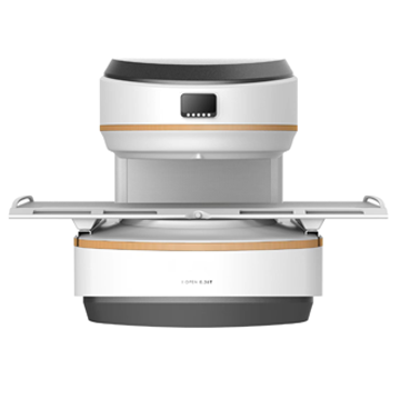 The 0.36T MRI system is a medical diagnostic magnetic resonance system. The main magnet of the system embodies the “C” shaped design type, single-pole structure. The structure has a greater degree of openness (opening angle greater than 270 degrees), which provides the patient with greater comfort. The main magnet has the advantage of compact design, small size (1.7m x 1.1m x 1.7m) and light weight (15T) with a static magnetic field strength of up to 0.36 Tesla.
Magnet
The MRI 0.36T has an open C-shaped magnet with active protection. The stability of the magnetic field is ensured using a patented magnet design and a specialized system for maintaining a constant magnet temperature.
The constant temperature magnetic system is composed of an insulating layer of magnetic insulation and an automatic temperature controller, which keeps the magnet temperature stable higher than the room temperature, which greatly improves the stability of the system by canceling the influence of room temperature fluctuations.
The fluctuation of the magnet field is small; The 5-G field is kept within 2.5 m or less in all directions, allowing the system to be installed in a small space. The complete system can be installed in a space of 22.5 m2.
Excellent magnetic field uniformity ensures excellent image quality with a 40cm field of view.
The open C-arm magnet design is based on a research and clinical foundation in full body imaging. The result is a high-resolution, wide-field magnet optimized for imaging different parts of the body.
• Field strength: 0.36T ± 5%
• Type: Permanent
• Field orientation: vertical
• Weight: 15000 kg
• Uniformity: 40 cm DSV 2.5 ppm.
• Gradient strength: 28.5 mT/m
• Slew rate: 95 mT/m/ms
• Aperture: 40 ± 1 cm
• Horizontal open angle: 300°
• Environment: Internal thermal control system
Patient table
A patient table is used to position the patient with appropriate RF receiving coils to the isocenter of the magnet for MR scanning.
The patient unit consists of two parts: the patient table and the positioning system. The patient table includes a table deck, deck carrier, cushions, etc.
The positioning system includes laser positioning and LED screen. Two LED screens display table coordinates simultaneously.
• Table width: 60 cm
• Table height: 76 cm from the floor
• Max. patient weight: 240 kg
Gradient system
The 0.36T MRI gradient system is state-of-the-art, capable of operating at 28.5 mT/ms with a slew rate of 95 mT/m/ms.
• Maximum voltage: 150 V
• Maximum current: 150 A
Higher gradient power maintains greater gradient strength, allowing the system to provide thinner slice thicknesses and smaller field of view minimums, and allows rapid (one-shot) acquisition of a highly compressed TSE sequence.
Digital RF System
A flat quadrature transmitting coil is built into the pole pieces, providing a strong, uniform field. The 6 kW RF power amplifier allows the use of short pulses, enabling fast image sequences.
Reception platform
The system is equipped with a fully digital two-channel hardware platform. This technology allows the use of phased array. These coils provide higher signal-to-noise ratio and better image quality.
The RF receiving system adapts quantum optical fiber transmission technology. Analog/digital conversion and quantum laser modulation are placed inside the magnetic room, which helps to significantly reduce signal loss and prevent interference during transmission. As a result, the image quality is effectively improved.
RF Receiver Coils
MRI 0.36T RF coils provide excellent signal to noise ratio due to the presence of preamplifiers in all coil elements and other receiver coils, ensuring proper signal amplification. The 0.36T MRI comes with a complete set of RF receiver coils to support a wide range of clinical applications from head to toe. Dedicated preset routines are available for all coils.
• Coil design: Phased coil
• Pick-up coils: Head
Neck
Shoulder
Knee
Body (large)
Body (medium)
Wrist
Flexible (optional)
Baby head coil (optional)
Chest coil (optional)
• Max. RF Power: 6KW
• Preamplifiers: Fully digital, magnetically fixed inside
• Positioning: Comfortable patient positioning and
restriction of movement.
Phased head coil
The phased head coil is optimized for imaging the head and upper cervical spine. The reel can be opened for quick and easy positioning.
Functions:
o Phased coil with high signal-to-noise ratio
o Large coverage for easy positioning.
o Split design for easy positioning
Phased neck coil
Coverage of the cervical coil is uniform from the cerebellum to the upper thoracic spine. The top of the reel can be removed for quick and easy positioning.
Functions:
o Coil phased with high signal-to-noise ratio
o High homogeneity from the cerebellum to the upper thoracic spine.
o Split design for easy positioning
Body coil phased
The Large Body/Spine Phased Coil has two elements that are suitable for imaging the chest, abdomen, pelvis, thoracic and lumbar spine in larger patients. The top of the reel can be removed for quick and easy positioning. In addition, the coil also slides under the patient for ease of positioning.
Functions:
o Phase array coil with high signal-to-noise ratio
o Split design and sliding support for easy positioning.
Shoulder coil phased
The phased shoulder coil has a special design that allows it to be worn on the shoulder. The spool loop slides along the arm for easy positioning.
Phased knee coil
The phase array knee coil is used for knee. The reel can be opened for quick and easy positioning.
Small joint coil (wrist)
Special design for wrist.
Flexible phased coil (optional)
The flexible coil is designed for obese patients and allows wider application of the i_Open 0.36T system.
Baby head coil (optional)
Special design for babies.
Computer control system
Server
Host computer with Intel CORE 2 DUO 3.5G processor or higher and at least 4GB RAM for general system management and processing, allowing simultaneous patient registration/pre-registration, MRI scanning and imaging, image reconstruction, image review, printing, etc. has significantly increased patient throughput. A 1TB hard drive is used for system software and data storage. CD/DVD RW for data archiving, backup and transfer.
o CPU: INTEL CORE DUO 3.5 GHz or higher
o Memory (RAM): 4 GB or more
o Hard Drive: 1TB or larger - Can store up to 6 million uncompressed 256x256 images.
o Archiving images: CD/DVD-RW
o Operating system: Windows 10
o Graphical interface: Windows based
o Network: DICOM 3.0 storage, media sharing, sending, printing and worklist
Operator console
i_Open 0.36T is designed to be operated by one operator. Patient administration and scan management, viewing and transfer of image data, image processing and printing, and system management can be performed using convenient and intuitive controls. Simple mouse clicks enable routine actions, providing high patient throughput and operator convenience. The operator console consists of:
o Microsoft Windows 10 operating system for efficiency and ease of operation.
o Desk with space for equipment and documents
o 24" TFT LCD monitor for professional picture quality
Patient's environment
i_Open 0.36T is the most convenient of the open scanners; Even the most anxious and claustrophobic patients will feel at ease. The C-arm magnet has a wide 40cm diameter opening for maximum patient access. The magnetic poles are small, which provides the patient with a complete view of the system from the outside in almost all studies.
o Patient communication system for two-way communication with the patient.
o A set of soft mattresses ensures patient comfort.
o Low noise system allows the patient to feel at ease during the entire scanning procedure.
o Manual nurse call button allows the patient to attract the attention of the operator without speaking (optional).
o The patient's music system will effectively relieve anxiety and emotional stress (optional).
Gating package (optional)
Breathing airlock
Respiratory gating can be used to reduce artifacts produced by breathing motion by collecting data based on the phase of the respiratory cycle. The device uses the difference in relative humidity between inhaled and exhaled air.
The respiratory gating package contains:
o balloon attached to the stomach for a pressure sensor
o pressure to digital signal converter
o Software displays breath waveform
o Trigger position can be selected by customer
Pulse strobe
The use of pulse triggering and gating is intended to monitor pulsating pulses.
Functions:
o wrist-mounted pressure sensor
o pressure to digital signal converter
o Software displays pulse waveform
Software Description
The software package is intended for use with magnetic resonance imaging systems i_Open 0.36T. The core software, called To-Station, will help operators and clinicians step-by-step patient enrollment, system setup, acquisition, processing, analysis and storage of 2D and 3D images, as well as integration of image enhancement, DICOM printing, etc.
Software package
o Scan management software
o Image reconstruction software
o Image processing software
o Image analysis software
o Image format conversion software (JPEG, BMP, etc.)
o Image viewing software
o System check software
o Real-time printing software (DICOM 3.0 standard)
o Software for remote maintenance
Extended capabilities
Patient pre-registration
Support for patient pre-registration during scanning without limiting the number of pre-registered patients improves operational efficiency.
9 fragments of scout images
9 slices of 3D survey images per pilot scan in 25 seconds give you 3 choices in each dimension, greatly improve scan positioning accuracy and ensure high patient throughput.
Scout line
The Scout's image can be displayed in the lower right corner and in the sidebar. This makes it very easy to know the exact position of the cut, which greatly improves the convenience of clinical diagnosis.
Image comparison
Provide comparison of multiple slices on one screen.
Smart Icon Management
Improve operator efficiency by customizing interface icons, repositioning each icon, and grouping icons.
Non-film capabilities
The VCD/DVD archive automatically integrates intelligent image viewing software called “to viewer”, which allows you to view archived images on any other PC.
Abundant sequence database
Provides free sequence updates.
The sequence database can be configured and classified by the operator. The system provides the operator with help information and automatically checks the correctness of the parameters.
Settings can be reset to default values
Standard DICOM 3.0 interface
- DICOM Modality Worklist (RIS Interface)
The Radiological Information System Interface option ensures automatic transfer of patient information from the hospital DICOM RIS to the MRI console operator console, thereby eliminating re-entry and possible errors.
Proper data transfer during the first step of MRI setup ensures data integrity and ensures proper communication between images and other patient data in the department information system or PACS. Functionality conforms to DICOM definitions.
It provides worklists for predefined time windows, sorted by time intervals and automatic transmission:
o Registration number
o Planned stage of the procedure
o Patient name
o Patient identification
o Patient gender
o Patient weight (if known in RIS)
o Name of referring physician
- DICOM Request/Receive Service Class as Provider
Supports database browsing from a DICOM workstation and sends copies of requested images in DICOM format.
The i_Open 0.36T DICOM Statement of Conformity contains complete information on the implementation of the DICOM standard.
Render Sequences and Parameters
Pulse sequences:
o Spin Echo Scout Image (3 x 3 scout images, each view has 3 scout images for orientation)
o Spin echo (SE)
o T1 weighted image with Spin Echo
o Proton Density Weighted Image from Spin Echo
o T2 Weighted Image with Fast Spin Echo
o Proton Density Weighted Image with Fast Spin Echo
o Dual contrast image with Fast Spin Echo
o 3D Fast Spin Echo
o Gradient echo (GE)
o T1-weighted image with gradient echo
o T2-weighted image with gradient echo
o Gradient echo with fast dephasing
o Fast rephasing of gradient echo
o Rapid breath-hold gradient echo dephasing
o 3D Fast dephase Gradient Echo
o Inversion recovery (IR)
o Fat suppression with Inversion Recovery
o Water suppression using inversion recovery
o Fat suppression with Fast Inversion Recovery (STIR) technology
o Water suppression with Fast Inversion Recovery (FLAIR) technology
o Heavy T1-weighted image with inversion recovery
o 2D/3D Time of Flight for angiography (TOF)
o Magnetic resonance cholangiopancreatography (MRCP)
o Magnetic resonance urography (MRU)
o Magnetic resonance myelography (MRM)
o Magnetization Transfer Contrast (MTC)
o Line scan diffusion weighted imaging (LSDWI)
Image reconstruction: 2D Fourier transform
3D Fourier transform
Half Scan
Cut thickness: 2D: from 0.5 mm to 10 mm in 0.1 mm increments
3-D: from 0.1 mm to 10 mm in 0.1 mm increments
Interslice Distance: Adjacent slices are available, system default is 10% interslice distance.
Cut orientation: transverse
Sagittal
coronal
Oblique and double oblique
Off center
Acquisition Matrix: 2-D: 128 x 128 to 256 x 256
3D: from 128 x 128 x 24 to 256 x 256 x 128
Field of view: 20 mm to 400 mm, 1 mm increments
Number of slices: 2-D: 1 – 256
3-D: 1–500 (maximum 50 slices x 10 blocks)
Image processing and manipulation
WDM "ToStation" provides powerful and easy-to-use tools for working with images.
Help: Offline Tutorial
Multitasking:
o All operations can be performed in parallel.
o Supports patient pre-registration for scanning without limiting the number of pre-registered patients, improving operational efficiency.
o Background task indicator
Queue for the exam:
o Manage and schedule a scan queue for a full survey
o Configuring protocols with archiving
Positioning:
o Visualize the current image with geometric references to the reconnaissance image.
o Graphical positioning using the mouse Image tools (on one image or on the entire series):
o Window width/level
o Scale
oh Pan
o Rotate
about the mirror
o measurements
o Distances
o ROI (manual, rectangular, oval), size, environment, standard deviation
o Image annotations
o Comparison of multi-slices on one screen
Database features:
o Search (alphabetically, chronologically, by patient ID, body part, gender, age, etc.)
o Sort
o Archiving and export functions to CD/DVD-ROM
CD/DVD Archiving: To ensure that images archived on CD/DVD can be viewed on any Windows PC, "ToViewer" - an intelligent image browser will be automatically burned to each disc.