Floor (graphy, scopy)

A universal two-person digital system for clinics and hospitals, private and public, with a high flow of patients. The system can meet the needs of most common examinations, both vertical and horizontal, including breast examinations. Actual contents and features vary depending on the configuration selected.
Peculiarities:
Adjusting the height of the tube with a stand
X-ray tube Siemens
Automatic exposure control
Radiation dose indicator (optional)
Two Flat Panel Detectors measuring 43x43 (graphy – detector for the table, graphy/copy – detector for the Buki stand)
Multispectral image processing
Branded workstation WD Acquire
Panoramic X-ray function (optional)
|
Name |
Specifications |
| High frequency generator | Catalog number: GFS501-1 Power: 65kW Radiography kV: 40~150Kv; mA Range: 10~800mA Fluoroscopy kV: 40~125kV, Fluoro mA: 10~40mA Exposure time: 1ms~ 10s, mAs: 0.1mAs~ 1000mAs Preset programs: 600 positions, Automatic exposure control Power: 380VAC, three-phase Network integration: generator parameter control and image acquisition are integrated on a single platform. Detection and diagnosis of faults. |
| Flat Panel Detector | Catalog number: WDF 4343RWiDetector type: a-SiEffective area: 430mm×430mm (17”×17”)Weight: 4.3kg Matrix (pixel): 3k ×3k, (9.4 megapixels), Pixel size: 139 umAnalog-Digital Converter: 16bit Spatial resolution: 3.4 lp/mm Image reconstruction time: <8s DQE: ≥70% MTF: ≥70% (@ 1.0 lp/mm) Ambient temperature: 10℃~40℃, Storage temperature: -15℃ ~ 55℃ Data transmission: WIFI / Giga Ethernet Control command transmission: WIFI / Giga Ethernet Calibration mode: offset calibration, gain calibration, error pixel calibration, line noise calibration Internal trigger control mode, applicable for upgrading the general system |
| Dynamic Flat Panel Detector | Catalog number: WDF 4343R (installed in Buki rack) Detector type: a-Si Size: 492mm×492mm×38mm Effective area: 43cm×43cm (17”×17”) Weight: 5.5kg Matrix (pixel): 3k ×3k, (9, 4 megapixels), Pixel size: 139 um A/D converter: 16bit Spatial resolution: 3.4 lp/mm Image reconstruction time: <3s Dynamic data acquisition rate: 25, 15, 4, 2, 1 fps Ambient temperature: 10℃~40℃, Storage temperature: -15℃~55℃DQE: ≥70%, MTF: ≥58% Data transmission: Giga Ethernet Control command transmission: Giga Ethernet Calibration mode: offset calibration, gain calibration, bad pixel calibration |
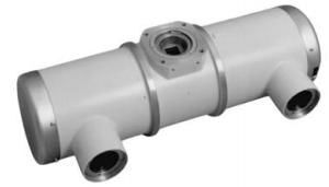
| X-ray tube assembly SIEMENS |
|
| Collimator | Catalog number: XS2-7 Radiation field (SID = 1000 mm): max.: 430 mm × 430 mm; Min:0mm×0mm Operating mode: Auto/manual |
|
Table & Buka stand |
X-ray table Catalog number: SC4-5 Height from table top to floor: ≤ 670 mm Tabletop movement range (floating) longitudinal ≥ 905 mm, transverse ≥ 260 mm Longitudinal tube movement: ≥ 2400 mm X-ray tube vertical movement (tube focus on floor): 525mm~1760mm (motorized) X-ray tube rotation along the horizontal arm: +120°~-120° Tube column rotation: 0°~±180° (fixed at ±90°, 0° and 180°) Longitudinal movement in relation to Beech post ≥ 540 mm Tabletop weakening equivalent <1.2 mm Al Load capacity: 250 kg LCD touch screen: 10.1 inches Beech Stance: Catalog number: LS-6 Detector vertical movement range (from center to floor): 370mm ~ 1800mm |
| Lattice | Mesh ratio: ≥10:1, f0: ≥100cm, Aluminum based |
| Remote control | Remote control of mechanical system movement |
| Imaging workstation | Hardware Configurations: Processor: Intel Core 2 Duo (Core 2 Duo) ≥3.7 GHz Memory: 4 GB; Hard Drive: ≥1TB DVD R/W System interface: USB, 1000 MB network interface, DVI/HDMI monitor 24-inch LCD monitor (color or monochrome); Resolution: 1920×1080 OS: Windows 7 or later Software Configuration: WD-ACQUIRE Image acquisition: setting acquisition conditions, setting mechanical position, APR Fluoro data acquisition: 25 fps, 15 fps, 4 fps, 2 fps, 1 fps Data management: patient registration, patient information and image management, report management Recording and displaying exposure data DICOM Service: Send, Receive, Store, Query, WORKLIST, MPPS, Validate, etc. DICOM SR: reporting, storage, printing, sending Data statistics: statistics of counting exposure, body part, number of patients, etc. for a specific date or period. RIS support. Image processing: window width/level, automatic window width/level adjustment, preview, preset window width/level, inverting positive and negative images; The image is flipped, rotated, scaled, moved; Image interpolation edge enhancement, original image annotation restoration, character/number annotation, image annotation, tape measurement, area measurement, auto-tuning. Image printing: DICOM printing, paper printing, manual selection of images displayed for printing, one-key annotated image printing, compatible with various printing equipment, film format printing, number of prints, print queue management, stop/start preset. Image enhancement filter: According to the different physiological structure of body parts and different diagnostic requests of doctors, and different clinical requirements, algorithms are optimized for different parts of the body. Personal settings: format and layout, default settings, toolbar settings. Other Features: Users can define the display format or layout; You can use different methods to view patient images and determine how to find information. Redundancy function, pixel optimization, diagnostic workstation image processing software interface in English, support high-speed lossless compression transmission, support online decompression. The operating interface is integrated with generator control and mechanical system status display, allowing all generator parameters such as kV exposure, mA, mAs, exposure time, ionization chamber activated field, APR, etc. to be configured on the operating interface. Exposure status and error code. |
 Catalog number: RAY-14_1