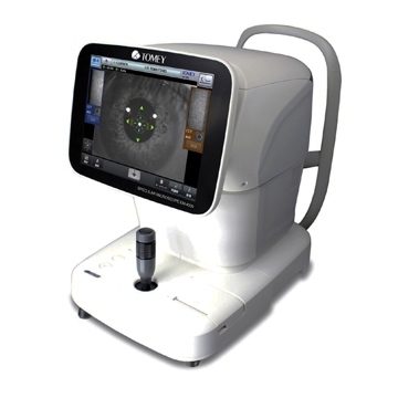
Endothelial microscope EM-4000
• Automatic targeting, measurement and analysis
• Built-in thermal printer
• Large 10.4" touch screen
• Increased number of fixation points
• Database maintenance
Defined parameters:
• Density and morphology of corneal endothelial cells
• Central corneal thickness
The principle of operation is based on photographing an enlarged image of the cornea of the eye, illuminated by a slit diagonally directed light source. Determination of corneal thickness occurs due to light reflected from the surface of the cornea and the endothelial layer of the cornea.
The EM-4000 uses reflected light for its positioning and autofocus system.
Automatic targeting and measurement
EM-4000 is ready to do almost everything yourself! Guidance and measurement are carried out automatically. Manual mode is available if necessary.
13 Measuring Areas and Automatic Pachymetry
The EM-4000 is capable of measuring a large area: the system automatically analyzes more than 300 corneal cells. Images can be obtained from 13 zones: central and 12 peripheral. And in addition, for each center measurement, pachymetry is also measured non-contactly!
Rapid automated endothelial cell analysis
The software gives access to all the necessary endothelial data: number and density of cells, morphology - polymegathism and pleomorphism. High quality images allow you to identify any abnormalities or degeneration of the endothelium. It is also possible to manually adjust the assessment of the corneal area.
Database and built-in printer
Thanks to the established database, two selected studies can be compared simultaneously: before and after surgery. Data for approximately 16,000 patients can be stored on an SD card installed in the main unit. Storing data in the device memory allows for additional analysis.
Indications for use
Inflammatory diseases of the cornea (keratitis), dystrophic diseases of the cornea (keratoconus, etc.), dry eye syndrome, assessment of the corneal endothelium after corneal surgery (through corneal transplantation, keratorefractive surgery), solving the issue of choosing tactics in cataract surgery.
The optical examination method eliminates direct contact with the patient and does not affect the examined diagnostic parameters - this significantly increases the accuracy of diagnosis and comfort for the patient.