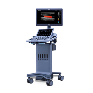
Digital Ultrasound Diagnostic System Acclarix LX3
New Generation
Features and flexibility
Combined with a compact design, advanced technology and intelligent workflow, Acclarix LX3 is designed to deliver more valuable innovation.
Its superior image quality plays an important role in prevention and early detection in primary care, enriching diagnostic applications and increasing clinical value.
System Features:
• 21.5” high resolution monitor with tempered glass coating
• 14” touch screen with customizable user interface
• Possibility of installing a gel heating device
• Ability to change height
• 5 active ports
• Battery for uninterrupted operation of the device
The EDAN Acclarix LX3 ultrasound scanner provides excellent image quality for ultrasound diagnostics in various areas:
• Abdominal examinations of adult and pediatric patients
• Obstetrics and gynecology, incl. 3D/4D
• Cardiology
• Vascular examination
• Examination of superficial organs
• Research of the musculoskeletal system
• Neonatology
• Urology
Specifications:
• Imaging modes: B, B/B, 4B, B/M, M
• CW - Continuous Wave Doppler
• PW (Pulse Wave Doppler) - Pulse Wave Doppler
• CDFI (Color Doppler Flow Imaging)
• PDI (Power Doppler Imaging) - Power Doppler
• Directional PDI (DPDI) – Directional Power Doppler
• Pulse Inversion Harmonic - tissue inverse harmonic
• 4D volumetric visualization mode in real time
• 3D volumetric reconstruction mode
• Panoramic scanning mode
• Trapezoidal scanning
• Console height adjustment
• THI - Tissue Harmonic
• TSI - Technology for recognizing the specificity of the tissues being studied
• Scanning depth (mm): up to 300
• Cine loop up to 12287 frames
• Internal archive for storing images (archiving images and cine loops to the built-in 512GB or 1TB hard drive or USB)
Intelligent Workflow
• eOptimized - one key optimization in B/Color/PW
• Automatic PW tracing - one key to automatic measurement
• High capacity storage device with USB transfer.
• eTouch - effective control of swipe gestures.
• E-learning - user-friendly learning software.
• Innovative user interface layout with precise separation of functions.
• Multilingual interface (Russian, English, German, French, Chinese)
• Software for research: abdominal, obstetric, gynecological, small organs, pediatrics, cardiology, urology, vascular, thyroid and mammary glands
• Auto Doppler spectrum tracing with auto measurements
• Built-in ports: VGA -1 pc., Video -1 pc., USB -6 pcs., DICOM 3.0 -1 pc., (optional), Ethernet port, printer control port, pedal connection port
• Power supply: 220 V mains operation
Sensors:
• Convex sensor C5-2XD (2-5 MHz / H2-5 MHz)
• Linear sensor L10-4D (4-9 MHz / H5-10 MHz)
• Endocavital sensor E8-4D (4-8 MHz / H5-8 MHz)
• Sector phased sensor P5-1XD (1-5 MHz / H2-5 MHz)
• Volumetric convex sensor C5-2MD (2-5 MHz / H3-5 MHz)
• Linear sensor L12-5D (5-11 MHz / H6-12 MHz)
• Linear sensor L17-7HD (7-15 MHz / H9-17 MHz)
• Linear sensor L17-7SD (7-15 MHz / H9-17 MHz)
• Microconvex sensor MC8‐4D (4-8 MHz / H4-8 MHz)