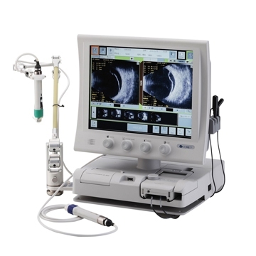
AB scanner UD-8000
The UD-8000 generates ultrasonic beams using software after digitizing the received waves - it is a digital radiation generator.
A digital radiation generator allows you to set focusing parameters (signal delay parameters for a ring matrix) more accurately than analog radiation generators. As a result, the resulting image has a higher resolution.
The physics of the propagation of ultrasonic vibrations is such that when moving from one medium to another, in relation to the carrier frequency of the radiation, side frequencies appear that are multiples of the frequency of the carrier vibration. For example, when the frequency of the carrier wave is 15 MHz, new radiation with a frequency of 30 MHz will appear at the input of the radiation receiver. A signal with a frequency of 30 MHz is called a secondary harmonic. Tissue Harmonic Imaging (THI) is a technique for acquiring images using only secondary harmonics.
Secondary harmonics have a number of advantages
Improved lateral image resolution due to narrow radiation;
Increased signal-to-noise ratio, reduced artifacts due to low secondary harmonic side lobes, etc.
Allows you to obtain high-resolution images with high noise immunity.
The intuitive touch screen control function, including video analysis function, offers you many options, such as: multiple image viewing modes (standard: 42 mm, wide: 54 mm), automatic video recording with continuous measurements (area, length, angle), as well as customized settings for each user.
The standard sensor is a multi-frequency B-sensor with the ability to change frequency from 15 MHz to 20 MHz within the measurement range.
The use of a UBM sensor makes it possible to visualize in detail the anterior part of the eye chamber, without violating the integrity of the eyeball, to carry out a qualitative and quantitative assessment of its structures, to clarify the spatial relationships of the cornea, ciliary body, iris, and lens in opaque refractive media, and to assess the condition of surgically created outflow tracts. The membrane waterproof cover (disposable) allows for quick and convenient examination without the use of gel. Thanks to this technology, you can examine the patient in a standing position.